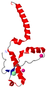
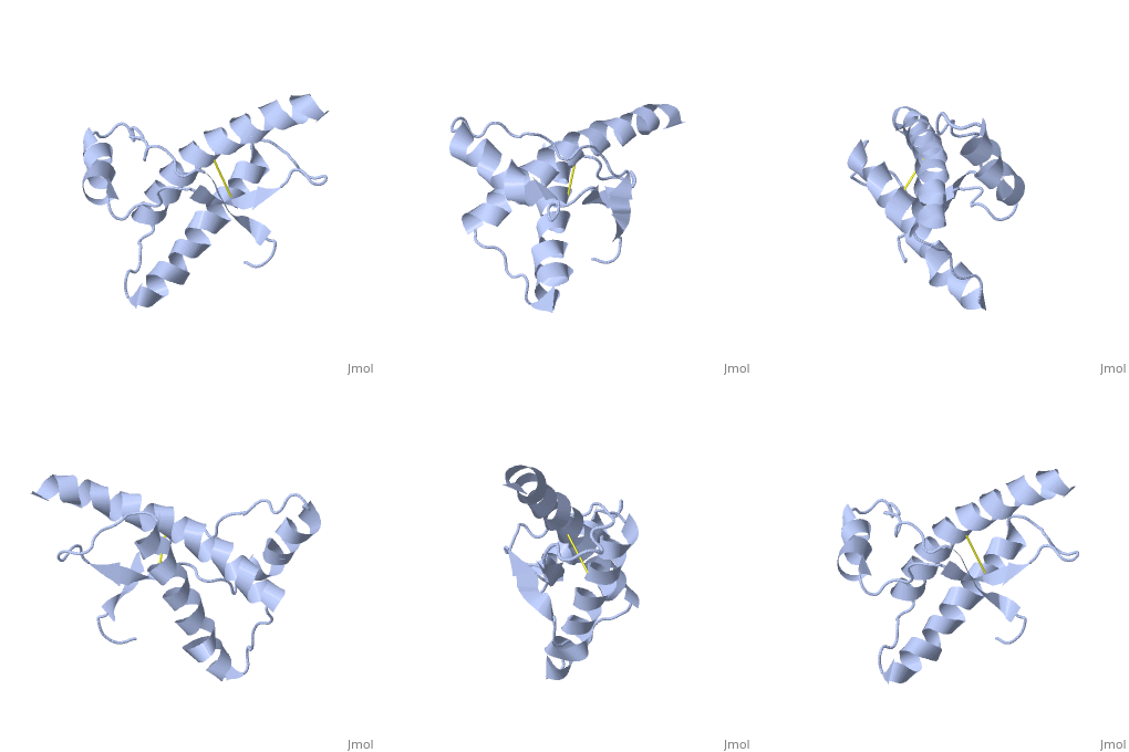
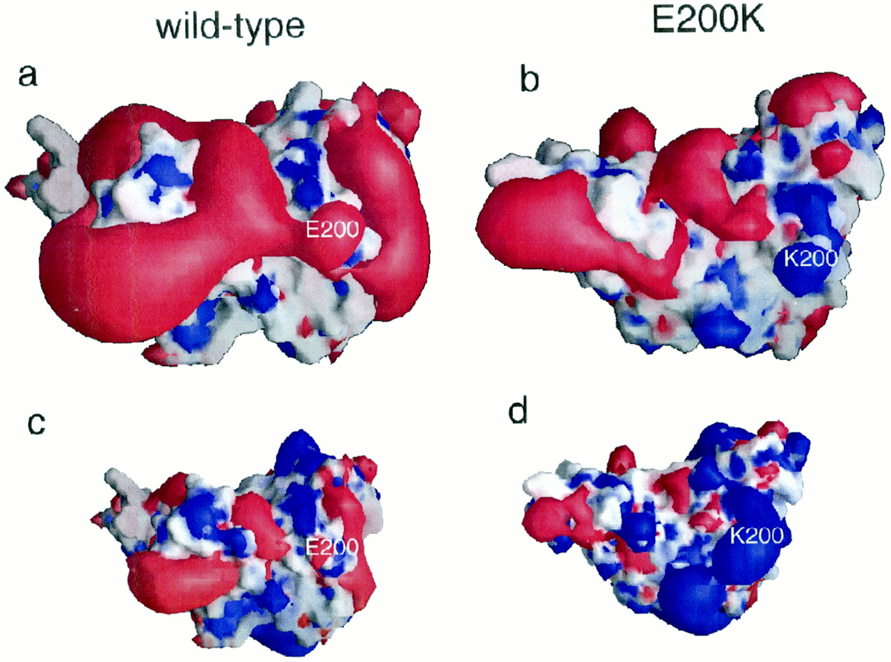
The , shown above, alter the function of Major Prion Protein's ability to re-fold, however their positions on the wild-type monomer and fully unfolded PrPSc, do not illustrate a clear mechanism for propagation. The dimer brings light to these residues influence on the infectious qualities of the disease causing residues.
It is theorized from this dimeric structure that the dimerization is the first step in amyloid formation and the presence of these dimers could possibly speed up the aggregation of PrPSc.
The following interactions and residue switching portray possible catalytic sites:
Residues 129, 200, and 164 to 170 are shown to exist right at the dimer interfaces (as noted in the previous structures). The location of these residues within the dimer indicates that their key operation is found somewhere in the dimerization process. [5][6][7]
Helix 1 (Ser 143−Tyr 157) exists at the . Many nonpolar residues form Van der Waals attractions. Acidic and mostly negative residues are shown in blue. Basic and mostly positive residues are shown in red. The interactions between these also stabilize the dimer interface.
Helix 2 (Asn 171−Thr 188) is to the C-terminal helix 3 (Thr 199−Tyr 225). Van der Waals forces are here between such nonpolar residues as valine, isoleucene, and nonpolar sections as histadine, methionine, and glutamic acid.[6]
The (residues 189-198) is where the actual unfolding has taken place.
More Van der Waals and electrostatic forces occur between the of the same molecule. These interactions would not be able to occur in the monomer.
between the dimers exist at Thr188 O−Gly195 N, Thr190 O−Lys194 N and Thr192 O−Thr192 N
between Asp 202 and Thr 199 stabilize the dimeric structure.
form a hydrogen bond located at the end of helix 3 and near the switch region of the other chain, which allow the inter-chain interactions to be specific.
Due to the importance of initial dimerization in the formation of prion aggregates, the step of dimerization presents a known step for treatments to target.[6]


