AF-Sm1
From Proteopedia

Structure
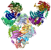
AF-Sm1, found the in the bacteria Archaeoglobus fulgidus is a member of a family called Sm and Sm-like (Lsm) proteins. This protein forms a heptameric structure and contains 1 α-helix, 5 β-sheets, and 5 loops between the α-helix and each of the β-sheets. Contained in the protein are two . The structure contains a predominate fold, called the Sm-fold which consists of an N-terminal helix followed by a sharply bent five-stranded β-sheet which leads to the protein having a barrel-type structure, commonly referred to as a “doughnut-shape.”[1] Uninterrupted hydrogen bonding occurs between β-sheets 4 and 5, leading to residues in loops 2, 3, and 5 being exposed at the inner surface of the ring and both the N and C termini being located at the outer surface of the ring.[1] A lysine residue located on loop 2 protrudes into the central cavity of the protein creating a positively charged region surrounding the central cavity on one side of the rings. [1] This protein has been linked due to its close structural resemblance, including sharing the Sm-fold structure, to the human dimeric D3/B and D1/D2 Sm complexes.[1]. See Transcription and RNA Processing.
Function
The function of AF-Sm1 protein is two-fold: eukaryotic and archaeal. In eukaryotes, this protein is involved in a range of RNA processing events, including pre-mRNA splicing, telomere replication, histone mRNA processing, and mRNA degradation. [1] They proteins form three distinct complexes with at least seven other Sm or Lsm proteins. Each complex interacts with a variety of RNAs, including the splicosomal U small nuclear (sn) RNAs, pre-RNase P RNA, telomerase RNA, viral RNA, and other RNAs. [1] A particular model of eukaryotic Sm protein function indicates that the Sm site RNA binds inside the doughnut-shaped ring involves a conserved residue near the 3 and 5 loops if the Sm proteins. [1] As for the archaeal function, much less is known about it when compared to the eukaryotic function. Recent crystal structure has revealed that the structure of the core domain of archaeal Sm proteins has been conserved over evolution. [1] Other parts of the protein have been highly conserved over time as well as they serve important functions structurally. AF-Sm1 in Archaea complexes with U5 oligonucleotide where oligouridine binds inside the heptomeric ring by specific hydrogen bonding to bases in pockets lined by highly conserved residues and by a specific stacking mechanism.[1] Based on that information, it is known that archaeal AF-Sm1 associates heavily with RNA rich in U-sequences.[1] It is thought that this protein plays a role in tRNA processing, perhaps with the mediation of other proteins or other RNA.[1]
3D structures of Sm proteins
- ↑ 1.00 1.01 1.02 1.03 1.04 1.05 1.06 1.07 1.08 1.09 1.10 Toro I, Thore S, Mayer C, Basquin J, Seraphin B, Suck D. RNA binding in an Sm core domain: X-ray structure and functional analysis of an archaeal Sm protein complex. EMBO J. 2001 May 1;20(9):2293-303. PMID:11331594 doi:10.1093/emboj/20.9.2293
| Please do NOT make changes to this Sandbox until after April 23, 2010. Sandboxes 151-200 are reserved until then for use by the Chemistry 307 class at UNBC taught by Prof. Andrea Gorrell. |
Proteopedia Page Contributors and Editors (what is this?)
Karly McLeod, Michal Harel, Jaime Prilusky, David Canner, Alexander Berchansky, Andrea Gorrell

