Image:180504 PurA Repeat 1 electron density.jpg
From Proteopedia
Size of this preview: 800 × 600 pixels
Full resolution (1687 × 1265 pixel, file size: 375 KB, MIME type: image/jpeg)
Summary
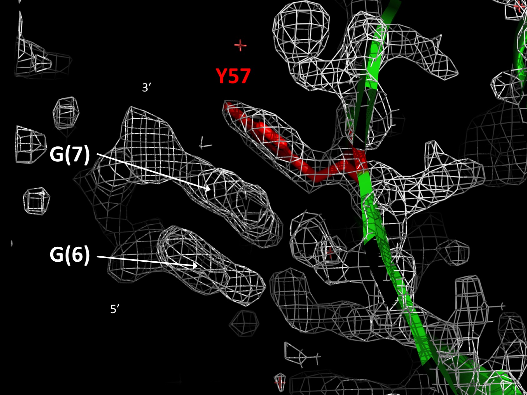
PUR repeat I electron density map showing base stacking between Y57 and guanine. The DNA strand present in this electron density map was omitted from the 5fgp PDB file but is shown in the original publication (Weber, J. et al., 2016, eLIFE).[1]
Licensing
|
|
File history
Click on a date/time to view the file as it appeared at that time.
| Date/Time | User | Dimensions | File size | Comment | |
|---|---|---|---|---|---|
| (current) | 20:53, 4 May 2018 | Andrea Foote (Talk | contribs) | 1687×1265 | 375 KB | PUR repeat I electron density map showing base stacking between Y57 and guanine. The DNA strand present in this electron density map was omitted from the 5fgp PDB file but is shown in the original publication (Weber, J. et al., 2016, eLIFE).<ref>PMID: |
- Edit this file using an external application
See the setup instructions for more information.
Links
The following pages link to this file:
Metadata
This file contains additional information, probably added from the digital camera or scanner used to create or digitize it. If the file has been modified from its original state, some details may not fully reflect the modified image.
| Horizontal resolution | 72 dpi |
|---|---|
| Vertical resolution | 72 dpi |
| Color space | sRGB |
